Background
General information about endometriosis
The uterus is a pear-shaped, hollow pelvic organ in which the foetus develops during gestation. In order to carry out this function, the uterus must demonstrate two characteristics: an adequate structural makeup and responsiveness to ovarian steroids (estrogen and progesterone). The uterus has a thick layer of smooth muscle called the myometrium and a thinner layer of columnar epithelium called the endometrium. The endometrium consists of a functional layer of cells, which is shed during each menstrual cycle and a basal layer from which the functional layer arises.
The endometrium has no other function than to act as a suitable medium in which a blastocyst can successfully implant after fecundation. The embryological origin of the endometrium as a part of an organ which arises from a very well-defined structure (the paramesonephric ducts) also makes it hard for such tissue to form directly in other places. It therefore follows that the only physiologically justified location of endometrial tissue is within the uterus. If one uses the term eutopic to refer to endometrium that exists in its expected location as the innermost layer of the uterus, one would consider the presence of ectopic endometrium (i.e. endometrium outside its normal location) as a pathological entity.
Accounts of the presence of ectopic endometrium first appeared in the 1860s when the Bohemian pathologist von Rokitanski analysed the contents of cystic, chocolate-coloured ovarian formations (6). These sporadic accounts and descriptions were eventually gathered into a systematic study at the beginning of the twentieth century by the gynaecologist John Sampson (6), who coined the term endometriosis to refer to the presence of ectopic endometrium in the pelvic cavity. Although it was largely passed over during the greatest part of the twentieth century, endometriosis started gaining new ground as a subject for scientific study with the increasing use of diagnostic and therapeutic laparoscopy in gynaecology. While the pathogenesis of endometriosis still remains unknown, modern studies have been focused on the molecular and cellular mechanisms by which this disease may come to exist.
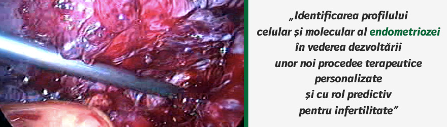
Endometriosis can therefore be defined as a benign disease of the female reproductive tract characterized by the presence of implants (or “islands”) of endometrial tissue outside the endometrial cavity. It is often a chronic, invalidating condition with two important manifestations - chronic pelvic pain and infertility.
Most sites of implantation involve the ovaries and Fallopian tubes, as well as the zones between the uterus and the anterior and posterior cul-de-sacs, the uterine ligaments and the pelvic wall (1). In other patients, the endometriotic implants spread beyond the inner genital organs and involve other organs in the peritoneal cavity, such as the intestines, the bladder or the lower parts of the ureters. Implants of ectopic endometrium have also been found in extragenital sites, such as surgical scars, the lungs or even the brain.
The prevalence of endometriosis is difficult to estimate in the general population, mainly due to the fact that the majority of women who seek treatment are aged between 15 and 49 years, with a peak of incidence between 28 and 35 years. These women are all of fertile age and the peak of incidence of endometriosis coincides with the peak of reproduction in women of a higher socioeconomic background. Cases of endometriosis in prepubertary girls or postmenopausal / posthysterectomized women have also been documented (1). However, they are infrequent enough to warrant the selective inclusion of fertile women in the various existent epidemiological studies concerning endometriosis.
A certain diagnosis of endometriosis is possible only after surgical excision and histopathologic analysis of specimens. One must also take into consideration that other women may be suffering from pauci – or asymptomatic forms of the disease and therefore never seek invasive treatment. The disease may eventually cause a significant decrease in the quality of life of the patient, but it is never a threat to her life. This is why screening programs which would possibly detect the aforementioned asymptomatic cases are justly deemed too costly.
Given these limitations, one can only estimate the prevalence of endometriosis to be between 5-10% of the general female population (7). Studies regarding women suffering from infertility show a far higher prevalence – it is estimated that 20 - 50% of infertile women have some form of endometriosis (8). Yet another study linked endometriosis to around 80% of cases of chronic pelvic pain in women of reproductive age (9). The link between the severity and extent of endometriosis and these two conditions is not very clear and will be discussed further on.
Although it is clearly a benign condition, endometriosis shares many characteristics with malignant pathology, particularly estrogen-dependent neoplasms. The ectopic endometrial cells demonstrate an aggressive potential which enables them to thrive outside their physiologic location due to:
- increased adherence to peritoneal cells (10)
- increased invasiveness due to a decrease in epithelial E-cadherin levels with concomitant increase in N-cadherin(11) expression and high secretion of metalloproteinases able to digest extracellular matrix proteins allowing cells to invade the collagen-rich basement membrane (4)
- increased capacity for neoangiogenesis (“inflammatory angiogenesis”)
- increased oxidative stress (12)
- resistance to apoptosis (13)
These characteristics are thought to be the result of somatically-acquired genetic alterations occurring mainly in advanced endometriosis states (14). It is important to note that, in spite of all these characteristics, endometriosis bears no relationship to endometrial cancer. It has, however, been associated with some forms of ovarian cancer, non-Hodgkin’s lymphoma and brain cancer (1).
Classic theories regarding the pathogenesis of endometriosis
Several theories regarding the pathogenesis of endometriosis have been put forth, but so far none of them has been able to account for all the features and variations of the disease. The most important four theories are: the presence of retrograde menstruation, caelomic metaplasia, Műllerianosis and lymphatic dissemination/transplantation (1).
A. Retrograde menstruation
Proposed by John Sampson in 1921 (6), this theory is the most widely accepted variant up to this date. Retrograde menstruation implies that a small quantity of menstrual blood flows backward through the Fallopian tubes and into the peritoneal cavity during each monthly cycle. Endometrial cells therefore become ectopic via this route and go on to implant themselves on various peritoneal surfaces and to form an endometriotic cyst. In recent years, retrograde menstruation has increasingly been documented as a physiological process that occurs in mammals and humans regardless of the presence of endometriosis. This means that there must be at least another factor present only in a minority of women that enables the endometrial cells to survive outside their normal environment.
B. Caelomic metaplasia
The caelomic theory states that peritoneal cells undergo metaplasia into endometrial cells. Peritoneal to endometrial metaplasia in a laboratory setting has been documented; however, this does not mean that it also takes place in vivo. Metaplasia is known to be a reversible process once the stimulus that triggered it ceases. There are no documented forms of “spontaneous regression” in endometriosis. Endometrial cells are in no way “hardier” or better equipped to withstand inflammation than peritoneal cells. Last but not least, metaplastic changes are associated with a higher risk of anaplasia (i.e. nuclear immaturity that leads to neoplastic changes), but endometriotic implants never evolve into endometrial adenocarcinomas.
C. Műllerianosis
Műllerianosis refers to the possibility that some primordial cells from the Műllerian ducts remain in an unaltered state (i.e. do not participate in organogenesis) and subsequently transform into endometrial cells independent of the newly-formed uterus. This theory would account for the frequency of pelvic endometriosis, as well as for the presence of endometriosis in non-menstruating females. Foetal autopsies have also brought evidence in support of this theory.
D. Lymphatic dissemination and/or transplantation
In order to explain the presence of endometriotic implants outside the peritoneal cavity, the dissemination of endometrial cells via lymphatic or vascular routes has been proposed. Iatrogenic transplantation of endometrial cells that went on to form endometriotic nodules has frequently been noted in Pfannenstiel scars (following gynaecologic procedures and caesarean sections) and even in the umbilicus (eponymously termed Villar’s nodule).
The diagnosis and staging of endometriosis
The diagnosis of endometriosis can either be incidental or the result of investigations for:
- chronic pelvic pain, which can either be cyclic or chronic, or take the form of dysmenorrhea, dyspareunia or defecatory pain (1), which is not correlated with the extent or severity of the disease, but may be related to the depth of invasion (16)
- infertility in the absence of genetic abnormalities, genital malformations, hormonal imbalances
The investigation methods employed in the diagnosis of endometriosis are varied and range from clinical examination and imaging (transvaginal ultrasonography, MRI) to diagnostic laparoscopy (1). Although current guidelines do not require histological evaluation for the diagnosis of endometriosis, some suggest that relying solely on laparoscopic findings in the absence of histological confirmation often results in overdiagnosis (15).
Current staging of the disease relies on the American Society of Reproductive Medicine (ASRM) updated classification (1997). It is a score based on the location of ectopic tissue, its extent and depth of invasion, the colour of lesions (signifying biochemical abnormalities concurrent with the activity of the disease) and the presence and consistency of peritoneal adhesions (15). It classifies endometriosis into four forms: minimal (score 1-5), mild (score 6-15), moderate (score 16-40) and severe (score > 40). However, it fairs poorly in estimating the fertile potential of the patient and the risks of recurrence, particularly in mild and lower-moderate forms of the disease (17).
Treatment options for endometriosis
Although some forms of endometriosis are asymptomatic (particularly in the initial stages of the disease), the large number of women that do suffer from its manifestations seek immediate long-term relief, often spending large amount of money on medical therapy, surgical procedures and assisted reproductive techniques and at the same time losing financial compensation due to frequent sick leave and decreased quality of work. Endometriosis must therefore be seen as an important public health issue with a great impact on the material and human resources of any state.
The medical treatment of endometriosis focused on the ectopic endometrium’s response to ovarian steroids. The endometrium (regardless of localization) responds to cyclic changes in steroid hormones by proliferation, differentiation and by the production of autocrine and paracrine factors(18). Estrogen stimulation induces hyperplasia and is accompanied by worsening of symptoms; therefore, medical therapies seek to inhibit estrogenic production or activity, mainly by inducing chronic anovulation.
Progestatives (taken orally or via af progesterone-releasing intrauterine device) have been used to antagonize estrogenic effects by causing endometrial atrophy and are relatively effective in diminishing pain and the frequency and extent of recurrences. Side effects include acne, weight gain and irregular menstruation. Selective progesterone receptor modulators (SPRM) are a new therapeutic alternative to progestatives and are presently undergoing clinical trials for their use in benign gynaecologic pathology (1).
Anovulation can also be induced by the use of androgens, which inhibit the midcycle luteinizing hormone (LH) surge, or gonadotropin-releasing hormone (GnRH) agonists, which inhibit the pituitary secretion of FSH and LH when they are administered in a non-pulsatile fashion. However, both types of compounds have significant side effects. The long-term use of GnRH agonists places the patient in an artificial menopausal state which has all the effects of hypoestrogenism, the most important one being the reduction of bone density leading to osteoporosis. Androgens lead to a phenotypical masculinization, characterized by hirsutism and voice deepening, accompanied by menopausal-like symptoms (hot flashes and anxious-depressive states) and dyslipidemia in some cases.
Recent studies have explored the role of aromatase inhibitors in the treatment of endometriosis (19), as it has been observed that the ectopic endometrium is capable of producing aromatase (an enzyme directly involved in estrogen synthesis) and thus generate its own source of hormonal stimulation. Aromatase inhibitors have side-effects similar to those of GnRH agonists and are currently being investigated as a therapeutic alternative for patients with severe endometriosis.
Surgical interventions in endometriosis constitute the most attractive option for the treatment of this disease, mainly due to the fact that laparoscopy is essential for the diagnosis of the disease. Laparoscopic excision of the lesions and lysis of peritoneal adhesions brings immediate relief of symptoms in most cases, but it does not prevent recurrences. Classical surgery (i.e. caeliotomy) is reserved only for cases with significant distortion of the pelvic anatomy, usually after multiple interventions for recurrences. In women over 50 with significant symptoms which find no therapeutic relief, hysterectomy and bilateral oophorectomy can be presented as a surgical option.
The therapy of endometriosis-associated infertility is also centred on surgery, which can eliminate adhesions and endometriotic cysts on the Fallopian tubes and ovaries and thus make fertilization, migration through a permeable tube and implantation more likely. In practice, however, many women are forced to resort to assisted reproductive techniques in order to achieve a pregnancy. Recurrences themselves as well as further adhesion formation following repeated surgical interventions for these recurrences may have a negative impact on fertility. The impact of long-term medically-induced endometrial atrophy and anovulation on the receptivity of the endometrium are also currently being studied.
The molecular and immune basis of endometriosis
The implantation and survival of ectopic endometrial cells as well as the clinical manifestations of endometriosis are probably the final link in a complex of aberrant biological processes. The molecular cause-and-effect cascade that leads to the formation, perpetuation and recurrence of endometriotic implants has been a subject of increasingly frequent exploration and debate. Most studies have arisen from what we currently know about the genetic, molecular, cellular and immunologic profile of the normal receptive endometrium of fertile mammals (including humans). This data has so far been compared to the characteristics of the same parameters in women suffering from endometriosis, mostly in an effort to understand the link between endometriosis and infertility. An important element of novelty characteristic of the proposed project is the comparison of samples of eutopic and ectopic endometrium prelevated from the same woman at diagnostic laparoscopy according to cellular and molecular biomarkers and the dynamic monitorization of the evolution of these findings after surgery and long-term medical treatment.
Research has so far been focused on:
- the genomic profile of patients suffering from endometriosis
- the role of epigenetic factors in the development of the disease
- the response of the ectopic endometrium to estrogen and progesterone
- possible immunologic dysfunction in patients with endometriosis
- the potential for invasion and angiogenesis of endometriotic cells
- peritoneal factors that may encourage the implantation of ectopic endometrium
A. The genomic profile of patients suffering from endometriosis
Some of the first studies linking genetic factors to endometriosis pointed out that the daughters and sisters of women with this disease are up to ten times more likely to suffer from it themselves during the course of their lives (20). Affected loci such as mutations at 10q26 and 20p13 have been described in an Australian cohort of over 1000 affected sisters (21), but this finding has not been independently confirmed. Currently, endometriosis is considered to be a polygenic inherited disease of complex multi-factorial aetiology. The first study of the gene expression profile of endometriosis using microarray technology was performed in 2002(22). Using a probe that contained > 4000 genes, researchers found eight up-regulated genes in endometriotic implants compared to normal uterine endometrium. There are genes currently known to be aberrantly expressed during the time of implantation in women with endometriosis, which may be candidates for the initiation and establishment of the disease or for implantation-based infertility.
B. Immunologic dysfunction in patients with endometriosis
The involvement of the immune system in the pathogenesis of endometriosis has been acknowledged in a number of studies published in the last decade. The majority of studies have focused on the presence and quantity of various cytokines in the peritoneal fluid of patients with endometriosis, secreted by increased numbers of activated macrophages. These macrophages have a prolonged stay in the peritoneal cavity, caused by the increased production of macrophage migration inhibitory factor (MIF) due to high levels of interleukin 1 (IL1) in the peritoneal fluid of women with endometriosis (23). Concentrations of tumor necrosis factor alpha (TNFα) and interleukin-6 (IL6) were also significantly higher in the peritoneal fluid of patients with endometriosis than in that of normal patients (24).
Recent focus has shifted towards substances which have a key role in both inflammation and implantation: interleukins 15 (IL15) and 18 (IL18). IL18 is a strong pleiotropic cytokine known to be involved in various immune diseases. Elevated levels of IL18 in the peritoneal fluid of endometriosis patients as opposed to non-endometriotic tissue were detected (25). However, more recent data contradict this finding and state that patients with severe endometriosis have higher IL12 levels irrespective of IL18 levels, suggesting that in this disease an alternative pathway is involved in induction of the T-helper 1 immune response (26). Another study assessed the expression of IL18 in eutopic and ectopic endometrium in patients with endometriosis by immunohistochemistry and semiquantitative RT-PCR. Women with endometriosis showed down-regulation of IL18 expression in ectopic and eutopic endometrium as compared with normal patients. We can conclude that our understanding regarding the variations found in IL18 expression is insufficient.
Ectopic endometrium expresses IL15 mRNA and protein with elevated levels compared with eutopic and control endometrium, irrespective of the phases of the menstrual cycle. Endometrial epithelial cells were found to be the primary site of IL15 expression. The peritoneal fluid content of IL15 shows a trend toward higher concentrations in women with endometriosis, compared to fertile control groups without pelvic pathology. Its elevated expression in ectopic endometrium suggests that this cytokine plays a key role in local inflammatory/immune responses that are critical in endometriosis-associated abnormalities (27). Peritoneal fluid IL15 levels were inversely correlated with the depth of invasion and disease stage, suggesting a possible role for this cytokine in the early pathogenesis of endometriosis. A gene expression profiling study on eutopic endometrial biopsy samples found IL15 up-regulated during the normal window of implantation, but significantly decreased in women with endometriosis(28).
C. The potential for invasion and angiogenesis of endometriotic cells
Ectopic endometrium cells are known to possess a high invasive potential. Hallmarks of this feature are a decrease in epithelial E-cadherin levels (reducing cell-to-cell interactions) and a synchronous increase in the levels of N-cadherin expression (mediating interaction with cells at ectopic sites) (11). Cytokeratin-positive and E-cadherin negative endometriotic cells have an invasive phenotype which is similar to that of carcinoma cells. High secretion of matrix-metalloproteinases (MMPs) allows the ectopic cells to digest the extracellular matrix and thus invade the collagen-rich basement membrane (4).
The link between endometriosis and infertility
Our current understanding of the association between endometriosis and infertility remains incomplete and is based on the differences between the expression of different markers in normal vs. endometriotic endometrium. Endometriosis is the second condition associated with implantation defects that can also result in lower in vitro fertilization success rates. Aberrant expression of endometrial biomarkers in endometriosis patients has been reported, suggesting that the endometrium is somehow affected by the inflammatory changes that accompany endometriosis.
Although several (adhesion molecules, cytokines, growth factors, lipids, etc) have been cited as having an important role in ensuring that implantation can take place, integrins are the best characterized markers of the receptive endometrium. The coordinated expression of integrins in both the embryo and the receptive endometrium suggests that they play a critical role during attachment and invasion. Recent evidence supports L-selectin, a member of the selectin family, as a key adhesion molecule during the initial attachment of the human embryo. The interaction between L-selectin and the carbohydrate moiety on the endometrial surface may serve as a bridge to bring the embryo into intimate contact with the endometrium, prior to firm attachment (29). The mechanism of cell adhesion is thought to be similar to the rolling actions of leukocytes on vascular endothelium as they first become tethered prior to more robust adhesion and invasion that involves integrin binding.
The pattern of expression of cytokines, growth factors and their receptors during the menstrual cycle suggests a role in both endometrial development and implantation. Recent findings support a role for IL-6 in the early pregnancy stages because endometrial mRNA is suppressed in the mid-secretory phase of patients with recurrent abortions (30). IL1α and IL1ra levels in the peritoneal fluid and serum of women with endometriosis were found to be higher than normal females. Impairment of regulation of the activity of IL1 in the peritoneal fluid and serum of women with endometriosis may therefore play an important role in the pathogenesis and development of endometriosis-associated infertility.
2. REFERENCES
1. Schorge JO, Schaefer JI et al – Williams’ Gynecology, First Edition - The McGraw Hill Companies, 2008 – Chapter 10 (“Endometriosis”): 225-43
2. Prentice A et al – Ovarian steroid receptor expression in endometriosis and in two potential parent epithelia: endometrium and peritoneal mesothelium -Hum Reprod. 1992 Oct;7(9): 1318-25
3. Cunningham DS, Hansen KA, Coddington, CC – Changes in T-cell regulation of responses to self antigens in women with pelvic endometriosis - Fertil Steril 1992 Jul; 58(1):114-9
4. Juhasz-Bőss I, Hofele A, Lattrich C, Buchholz S, Ortmann O, Malik E – Matrix metalloproteinases messenger RNA expression in human endometriosis grafts cultured on a chicken chorioallantoic membrane - Fertil Steril 2010 Jun; 94(1):40-5
5. Liu H, Lang JH – Is abnormal eutopic endometrium the cause of endometriosis? The role of eutopic endometrium in pathogenesis of endometriosis - Med Sci Monit 2011 Apr; 17(4):RA92-9
6. Benagiano G, Brosens I - The history of endometriosis: identifying the disease - Hum Reprod 1991 Aug;6(7):963-8
7. Wheeler JM – Epidemiology of endometriosis-associated infertility – J Reprod Med Jan 1989;34(1): 41-6
8. Strathy, JH, Molgaard CA, Coulam CB, Melton LH – Endometriosis and infertility: a laparoscopic study of endometriosis among fertile and infertile women – Fertil Steril Dec 1982; 38(6):667-72
9. Carter, JE – Combined hysteroscopic and laparoscopic findings in patients with chronic pelvic pain – J Am Assoc Gynecol Laparosc Nov 1994;2(1):43-7
10. Griffith JS, Liu YG, Tekmal RR, Binkley PA, Holden AE, Schenken RS – Menstrual endometrial cells from women with endometriosis demonstrate increased adherence to peritoneal cells and increased expression of CD44 splice variants – Fertil Steril 2010 Apr;93(6):1745-9
11. Starzinski-Powitz A, Handrow-Metzmacher H, Kotzian S – The putative role of cell adhesion molecules in endometriosis: can we learn from tumor metastasis? – Mol Med Today, 1999 Jul;5(7):304-9
12. Carvalho L, Podgaec S, Bellodi-Privato M, Falcone T, Abrao, MS – Role of eutopic endometrium in pelvic endometriosis – J Minim Invasive Gynecol. 2011 Jul-Aug;18(4):419-27
13. Nasu K, Yuge A, Tsuno A, Nishida M, Narahara H – Involvement of resistance to apoptosis in the pathogenesis of endometriosis – Histol Histopathol. 2009 Sep;24(9):118-92
14. Fauser BCJM, Diedrich K, Bouchard P et al – Contemporary genetic technologies and female reproduction – Hum Reprod Update 2011 Nov;17(6): 829-47
15. American Society for Reproductive Medicine - Revised American Society for Reproductive Medicine classification of endometriosis - 1996. Fertil Steril 67:817, 1997
16. Koninckx PR, Braet P, Kennedy SH, et al - Dioxin pollution and endometriosis in Belgium - Hum Reprod, 9:1001, 1994
17. Guzick DS, Silliman NP et al - Prediction of pregnancy in infertile women based on the American Society for Reproductive Medicine's revised classification of endometriosis - Fertil Steril 67:822, 1997
18. Vinatier D, Orazi G, Cosson M, Dufour P - heories of endometriosis - Eur J Obstet Gynecol Reprod Biol 2001 96:21–34
19. Amsterdam LL, Gentry W, Jobanputra S, et al - Anastrazole and oral contraceptives: a novel treatment for endometriosis - Fertil Steril 84:300, 2005
20. Eskenazi B, Warner ML – Epidemiology of endometriosis – Obstet Gynecol Clin North Am. Jun 1997;24(2):235-58
21. Treloar SA et al – Genomewide Linkage Study in 1,176 Affected Sister Pair Families Identifies a Significant Susceptibility Locus for Endometriosis on Chromosone 10q26 – Am J Hum Genet. 2005 Sep; 77(3): 365-76
22. Eyster KM, Boles AL, Brannian JD, Hansen KA – DNA microarray of gene expression markers of endometriosis – Fertil Steril 2002 Jan; 77(1):38-42
23. Kats R, Metz CN, Akoum A – Macrophage Migration Inhibitory Factor Is Markedly Expressed in Active and Early-Stage Endometriotic Lesions – J Clin Endocrinol Metab 2002 Feb;87(2):883-9
24. Iwabe T, Harada T, Terakawa N – Role of cytokines in endometriosis-associated infertility – Gynecol Obstet Invest 2002;53 Suppl 1:19-25
25. Oku H et al – Role of IL18 in pathogenesis of endometriosis – Hum Reprod 2004 19(3): 709-14
26. Fairbanks F, Abrão MS, Podgaec S, Dias JA, de Oliviera RM, Rizzo LV – IL12 but not IL18 is associated with severe endometriosis – Fertil Steril 2009 Feb; 91(2):320-4
27. Chegini N, Roberts M, Ripps B – Differential expression of IL13 and IL15 in ectopic and eutopic endometrium of women with endometriosis and normal fertile women – Am J Reprod Immunol 2003 Feb;49(2):75-83
28. Kao LC, Germeyer A et al – Expression Profiling of Endometrium from Women with Endometriosis Reveals Candidate for Disease-Based Implantation Failure and Infertility – Endocrinology 2003 Jul 144(7) 2870-2881
29. Alon R, Rosen S – Rolling on N-linked glycans: a new way to present L-selectin binding sites – Nature Immunology 2007 8, 339-41
30. von Wolff, M, Stieger S, Lumpp K et al – Endometrial IL6 in vitro is not regulated directly by female steroid hormones, but by pro-inflammatory CK and hypoxia – Mol Hum Reprod 2002 1096-02